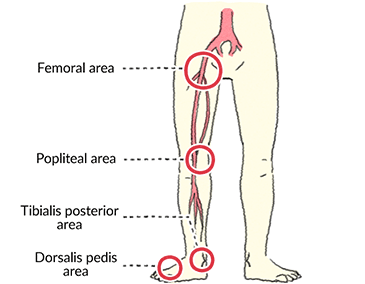
- Peripheral Pulses +3
- Peripheral Pulses 3+
- Peripheral Pulses 2+
- Peripheral Pulses Scale
- Peripheral Pulses +2
Thus, examination of the carotid pulse provides the most accurate representation of changes in the central aortic pulse. The brachial arterial pulse is examined to assess the volume and consistency of the peripheral vessels. UNEQUAL OR DELAYED PULSES. Inequality in the amplitude of the peripheral pulses. It may occur in one or both legs depending on the location of the clogged or narrowed artery. Other symptoms of PVD may include: Changes in the skin, including decreased skin temperature, or thin, brittle, shiny skin on the legs and feet. Weak pulses in the. Physical examination of the man.For more videos go to http://medicine.forumfree.it/?f=9837306.
Professional Reference articles are designed for health professionals to use. They are written by UK doctors and based on research evidence, UK and European Guidelines. You may find one of our health articles more useful.
Treatment of almost all medical conditions has been affected by the COVID-19 pandemic. NICE has issued rapid update guidelines in relation to many of these. This guidance is changing frequently. Please visit https://www.nice.org.uk/covid-19 to see if there is temporary guidance issued by NICE in relation to the management of this condition, which may vary from the information given below.
Pulse Examination
In this article
It is very easy to overlook the art of clinical examination when new technology can so easily be employed to make diagnoses. Systematic cardiovascular examination can provide a diagnosis quickly without need for invasive or expensive tests. Such routine examination can reveal an unexpected and timely diagnosis.
Trending Articles
Historically, in the Middle or Far East, doctors were expected to make many diagnoses on examination of the radial pulse alone. Still today, thorough examination of the pulse can provide a lot of information and help form an accurate diagnosis.

It is important to develop a reliable routine for examining the pulse and to refine and improve the technique throughout a career.
History
As with all clinical examination, there are aspects of the history which are particularly relevant to abnormalities in the pulse. There are many symptoms which may be relevant; however, some examples include:

- Cardiovascular symptoms including:
- Chest pain (at rest or on exertion).
- Palpitations.
- Syncope.
- Claudication.
- Tiredness or lethargy.
- Shortness of breath on exertion.
- Age (affects the likelihood of atherosclerosis).
- Past medical history (particularly of thyroid, cardiovascular and cerebrovascular disease).
- Lifestyle and occupation (the fit and trained athletes have very low pulse rates).
- Medication (many drugs can affect the pulse, including beta-blockers and digoxin).
Examination
General inspection
Observe the patient whilst taking the history. Look for:
- Apprehension or anxiety.
- Breathlessness.
- Cyanosis.
- Pallor or anaemia.
- Features of specific diseases, especially those associated with cardiovascular disease:
- Thyroid disease.
- Recognisable syndromes.
- Pulsations in the neck:
- Arterial and venous pulsations (jugular venous pulse) may be visible in the neck.
- Head movement:
- de Musset's sign (the head nods in time with the heartbeat - seen rarely in severe aortic regurgitation).
- Take the patient's hand and assess:
- Warmth (fever, thyrotoxicosis).
- Sweating.
- Tremor.
- Nails:
- There may be peripheral cyanosis.
- Clubbing of the nails (may indicate other disease, although clubbing can be congenital and benign).
- Splinter haemorrhages (infective endocarditis).
- Koilonychia (might indicate iron deficiency).
- Quincke's sign is pulsation of the capillary nail bed (with the very wide pulse pressure of aortic regurgitation).
Examining the pulse
Arterial pulses can be examined at various sites around the body. Systematic examination normally involves palpating in turn radial, brachial, carotid, femoral and other distal pulses. Palpation of the abdominal aorta would also form part of this systematic examination (to identify abdominal aortic aneurysms for example). Other sites may be examined for pulses, in special circumstances - for example, the temporal artery (for tenderness in temporal arteritis) and the ulnar artery (if the radial cannot be felt or before arterial access at the radial site).
Which pulse should be examined?
- This will depend on circumstances and whether there are specific clinical reasons for examining a particular pulse or for systematically examining all arterial pulses.
- In clinical examinations it is important that the student follow instructions and take clues from the questions posed by the examiner and the type of examination (distinguish, for example, between a request to examine a pulse and conducting a cardiovascular examination). Such examination technique can be practised according to the requirements of the particular examination to ensure success.
- Clinically it is traditional to examine the radial pulse first. However, there is much to commend routinely following this with examination of the larger brachial and carotid arteries to feel the nature of the wall and particularly the character of the pulse.
- There are, of course, specific reasons to examine all the pulses at different sites as part of a complete and systematic cardiovascular examination. As ever, in clinical practice there will be some selectivity to save time in the consultation.
| Systematic examination of pulses |
|---|
| Where and how? | 1. Radial artery | - To assess rate and rhythm.
- Simultaneously with femoral to detect delay.
- Not good for pulse character.
|
- Medial border of humerus at elbow medial to biceps tendon.
- Either with thumb of examiner's right hand or index and middle of left hand.
| 3. Carotid artery | Press back to feel carotid artery against precervical muscles.Alternatively from behind, curling fingers around side of neck. | 4. Femoral artery | - To assess cardiac output.
- To detect radiofemoral delay.
- To assess peripheral vascular disease.
|
- Deep within the popliteal fossa.
- Compress against posterior of distal femur with knee slightly flexed.
| 6. Dorsalis pedis (DP) and tibialis posterior (TP) arteries (foot) | |
- With the flat of the hand per abdomen, as body habitus allows.
| Do not press too hard for fear of obliterating the pulse.Establish whether the wall feels soft and pliable or hard and sclerotic.Identify the qualities or characteristics of the pulse by asking:- What is the pulse rate?
- What is the pulse rhythm?
- What is the character of the pulse?
What is the pulse rate?- A normal pulse rate after a period of rest is between 60 and 80 beats per minute (bpm). It is faster in children. However, if tachycardia is defined as a pulse rate in excess of 100 bpm and bradycardia is less than 60 bpm then between 60 and 100 bpm must be seen as normal.
- An irregular pulse or a slow pulse should be measured over a longer time. As a guide, it is unwise to measure a regular rate for less than 20 seconds (30 seconds being preferable) and an irregular pulse should not be measured over less than 30 seconds, preferably a full minute.
- Bradycardia may be physiological in a trained athlete, even if training was many years ago.
- Paroxysmal tachycardia can last a few minutes to several hours. It might be too transient to allow an ECG recording during an attack so that only clinical examination is available.
- Paroxysmal atrial fibrillation (AF) or, more rarely, atrial flutter, produces a ventricular rate that is dependent upon the refractory time of the A-V node.
- Re-entry tachycardia such as Wolff-Parkinson-White syndrome can produce a very fast rate in the region of 200 bpm.
- As a general rule, supraventricular tachycardia (SVT) produces a rate above 160 bpm and ventricular tachycardia (VT) below 160 bpm.
- Even in young people, very fast rates of 200 bpm or more can precipitate heart failure.
Peripheral Pulses +3What is the pulse rhythm?- Sinus arrhythmia occurs when there is variation of rate with breathing. It accelerates a little on inspiration and slows a little on expiration. This can be quite marked in children and adolescents but is uncommon over the age of 30. It can persist a little longer in the physically fit.
- Pulsus paradoxus:
- The pulse slows on inspiration in pulsus paradoxus and it can occur with pericardial effusion, constrictive pericarditis and severe pneumothorax, especially tension pneumothorax, severe asthma and severe chronic obstructive pulmonary disease (COPD).[1]
- In normal circumstances, the systolic blood pressure often falls slightly, by less than 10 mm Hg on inspiration; however, in pulsus paradoxus it falls by more than this.[2]This fall can be used to assess the severity of cardiac tamponade.
- Irregularity is more difficult to discern if the rate is fast.
- Note if it is regularly irregular of irregularly irregular:
- Variable heart block or premature ventricular excitation will cause either an extra beat or a missed one. Premature ventricular contraction may cause a missed beat because the ventricle has not had time to fill adequately and so the stroke volume is low. The beat following a missed beat, whether due to premature excitation or failure of the ventricle to beat, may be rather stronger than the others, as the ventricle has filled more in the longer diastole. This irregularity will follow a regular pattern.
- A much more random irregularity is a feature of AF. If the rate is fast in AF, it may be difficult to note if the irregularity is random or even if there is irregularity at all. It may be helpful to measure the rate at both the cardiac apex and the wrist and in AF there is usually a deficit at the radial pulse. This is usually done with two people timing simultaneously but it can be done alone, not timing but merely noting if the rates differ. The rate in AF and the rarer atrial flutter depends upon the degree of A-V block but it can be very fast.
- It has been suggested that a way to distinguish between causes of irregularity is to get the patient to exercise to increase the pulse rate. In premature ventricular excitation it will reduce or disappear. In AF it will increase the irregularity or at least not reduce it.
- Currently, most clinicians would use the ECG for a more reliable means of distinction.
What is the character of the pulse?Finally, note the character of the pulse. This incorporates an assessment of the pulse volume (the movement imparted to the finger by the pulse) and what has been described as the 'form of the pulse wave'. The pulse character must be interpreted in the light of pulse rate. - Cardiac output is the product of stroke volume and heart rate. Thus, a slow pulse may be associated with a high stroke volume and, as there is a long time between each ejection, the pulse pressure (the difference between systolic and diastolic pressure) will be high.
- In shock, the pulse will be fast but weak. This might be from hypovolaemia or cardiogenic. In congestive heart failure one of the first features is tachycardia.
- A hyperdynamic circulation occurs in emotion, heat, exercise, anxiety, pregnancy, fever, anaemia and thyrotoxicosis. The pulse rate is raised but the pulse is full and bounding. Cardiac output is high and peripheral resistance is low.
- Disease of the aortic valve will affect the nature of the pulse wave:
- In aortic stenosis, the wave is slow to rise and the pattern of the arterial pressure is rather flat - the slow rising pulse.
- In aortic regurgitation, the stroke volume is high because a significant amount of blood sinks back into the ventricle and has to be pumped again. Furthermore, the incompetent valve will let the arterial pressure fall markedly in diastole. Hence, a bounding, dynamic pulse collapses to give a very wide pulse pressure. This is called a collapsing or water hammer pulse. The water hammer is a piece of Victorian engineering that is rarely seen these days. The collapse of the pulse pressure can be felt with even greater effect if the patient's hand is raised over his or her head so that the radial artery is palpated at a level rather above the heart. Severe aortic regurgitation, classical of syphilitic aortitis, can cause the head to jerk with each pulse (de Musset's sign).
- In mild aortic stenosis with reflux, the pulse detected may have two peaks as well as being slow rising. This is the so called bisferiens pulse.
Studies have correlated markers of arterial stiffness (eg, pulse-wave velocity and pulse pressure) with risk for the development of fatal and non-fatal cardiovascular events.[3, 4]  The next stepPeripheral Pulses 3+This systematic examination of the pulse will give a great deal of information. Examination of the rest of the cardiovascular system should give a very clear idea of the diagnosis or at least put the examiner in a position to make a rational request for further investigations. Systematic examination of the pulse remains an essential part of clinical practice. Become a COVID-19 treatment pioneer today. Khasnis A, Lokhandwala Y; Clinical signs in medicine: pulsus paradoxus. J Postgrad Med 200248:46-9 Hamzaoui O, Monnet X, Teboul JL; Pulsus paradoxus. Eur Respir J. 2013 Dec42(6):1696-705. doi: 10.1183/09031936.00138912. Epub 2012 Dec 6. Liao J, Farmer J; Arterial stiffness as a risk factor for coronary artery disease. Curr Atheroscler Rep. 2014 Feb16(2):387. doi: 10.1007/s11883-013-0387-8. Boutouyrie P, Fliser D, Goldsmith D, et al; Assessment of arterial stiffness for clinical and epidemiological studies: methodological considerations for validation and entry into the European Renal and Cardiovascular Medicine registry. Nephrol Dial Transplant. 2014 Feb29(2):232-9. doi: 10.1093/ndt/gft309. Epub 2013 Sep 30.
Peripheral Pulses 2+I have my regular 8 month visit coming up with my cardiologist soon. Have had a ATA for over 5 years with little to no growth(around 5.3 cm). I've told when the time comes, the only treatment is... Health ToolsPeripheral Pulses ScaleFeeling unwell?Peripheral Pulses +2Assess your symptoms online with our free symptom checker.

|


Comments are closed.